
3-dimensional x-ray DVT
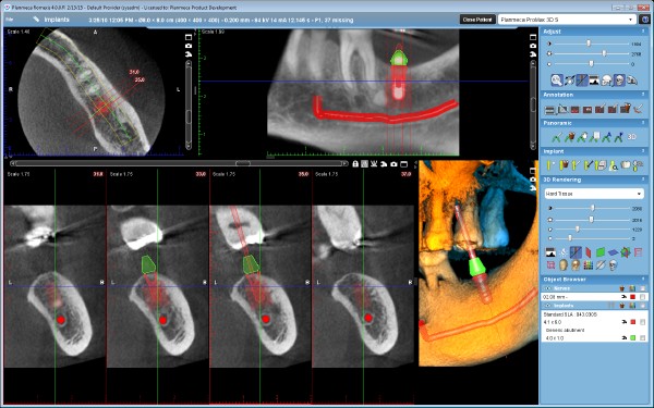
This is a three-dimensional imaging procedure (X-ray) which exposes the patient to much less radiation than, for example, a computer tomograph (CT), thanks to the latest technology. The radiation dose can also be reduced by restricting the field of vision. With the “ultra low dose” protocol of our device, the effective patient dose values of 2-dimensional devices are reached or partially undercut!
The DVT is used when conventional two-dimensional X-ray images do not provide enough information for a diagnosis or to precisely plan an intervention.
Examples:
- determination of the positional relationship of nerves or paranasal sinuses to wisdom teeth or other displaced/retained teeth/ planning of their removal
- presentation of the anatomy of root canals with complex root treatments or revisions of root treatments
- dental implant planning (increased safety, accurate navigation)
- presentation of temporomandibular joints and maxillary sinuses
- presentation of jaw and tooth fractures
- presentation of other pathological changes (tumours etc.)
Your advantages as a patient:
- best 3-dimensional imaging, thus greater reliability in diagnosis and treatment without additional referral
- quicker decision regarding the choice of therapy and thus prevention of major damage
- increased safety during surgical procedures thanks to precise advance planning
- low radiation dose despite 3-dimensional imaging
We look forward to explaining the new technology to you personally in our practice!
Useful Information for You
Questions About 3D X-rays
X-rays are ionizing radiation and are therefore potentially harmful, especially to rapidly regenerating tissues. For this reason, the benefits of an X-ray must be weighed against the risks. As much as necessary, as little as possible.
It is important to select the appropriate imaging technique for the specific clinical question and to implement the necessary protective measures. Pregnant women in their first trimester should not undergo X-rays. In the case of children, too, careful consideration should be given to whether an X-ray is indicated.
The following principle applies: the ALARA principle—as low as reasonably achievable.
However, when you consider that a panoramic X-ray exposes you to roughly the same amount of radiation as one hour in heavy traffic, and a digital volume tomography (3D X-ray) to about the same as a transatlantic flight, there is no need to fear these diagnostic methods. We are also exposed daily to natural radiation from space, the Earth, and building materials. This natural radiation is equivalent to approximately 400 dental X-rays per year.
Because modern digital X-ray systems use more sensitive sensors, the radiation dose can be significantly reduced compared to older analog systems.
During the first trimester, X-rays should be avoided whenever possible, as the risk of harm cannot be completely ruled out. After that, an X-ray may be justified in urgent cases (benefit vs. risk).
The anesthetics we use are safe for pregnant and breastfeeding women and pose no risk to the child. In most cases, we try to reschedule any necessary procedures until after the birth.